Amyloid β-protein and Synaptophysin

Amyloid β-peptide is normally produced in the brain from the amyloid precursor protein (APP). The amyloid cascade hypothesis of Alzheimer disease says that abnormal accumulation in the brain of amyloid β-peptide () causes disruption of synaptic neurotransmission and, eventually, the death of neurons and Alzheimer disease (see the image below).

Amyloid Cascade Hypothesis

During the past 20 years there has been a large amount of research into the normal physiological role of APP and the idea that  can disrupt synaptic neurotransmission.

In a previous blog post I mentioned that the WNT signaling pathway has been implicated in memory storage. There is evidence that the enzyme glycogen synthase kinase 3β (GSK-3β) is involved in memory deficits seen in Alzheimer disease.

A recently published article provides evidence that 42 binds to the synaptic vesicle protein synaptophysin42 disrupts the complex that is normally formed between synaptophysin and VAMP2 altering the way that neurons handle their synaptic vesicles.

If Aβ42 is involved in synaptic transmission by modulating several pre- and postsynaptic mechanisms then such research can identify potential targets for pharmacological intervention against age-associated memory loss.

In a previous blog post I mentioned the synaptic vesicle protein synaptogyrin-3. Synaptophysins and synaptogyrins are members of the tetraspan
vesicle membrane protein family and constitute major secretory vessicle proteins that bind to many other vessicle-associated proteins.

Related reading: The Alzheimer’s amyloid-degrading peptidase, neprilysin: can we control it?

Posted in Alzheimer disease, synaptic vesicles | Tagged , , , | Leave a comment

Regulation of Monoamine Oxidase in Parkinson Disease

Parkin regulated protein turnover.

In an earlier blog post I linked to an article about the potential role of dopamine metabolism and oxidative stress in Parkinson disease. Now the research group of Jian Feng reports that the protein Parkin regulates the expression of monoamine oxidase in human dopaminergic neurons (Parkin controls dopamine utilization in human midbrain dopaminergic neurons derived from induced pluripotent stem cells).

As early as 2003, experiments in mice linked low levels of Parkin to increased metabolism of dopamine by monoamine oxidase (1). One of the existing treatments for Parkinson Disease is inhibition of monoamine oxidase (see: “The role of rasagiline in the treatment of Parkinson’s disease“).

Feng’s group started with skin cells from human patients with mutated Parkin genes. They made pluripotent stem cells from the skin cells and then induced the stem cells to differentiate into dopaminergic neurons for use in their studies of the role of Parkin in human neurons.

Parkinson Disease patients with defective Parkin genes might particularly benefit from treatment with monoamine oxidase inhibitors.

Image source: European Iron Club.

Posted in L-DOPA, Parkinson disease, proteolysis | Tagged , , | Leave a comment

ProfCast

I finally used ProfCast software to add audio tracks to PowerPoint presentations.

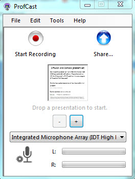
screenshot of ProfCast interface
ProfCast recording interface

This image (to the right) shows the ProfCast interface after drag/drop of the “Diffusion and Osmosis problem 1” PowerPoint presentation into the ProfCast window. Here is a link to the PowerPoint pptx file:

Diffusion and Osmosis problem 1

While you use ProfCast to record audio for the presentation, it creates PNG image files for each slide in the PowerPoint presentation.

Slide count and status

Slide count and status

ProfCast will also save an audio file. The audio format is ADTS. You can save your combined images and audio as an m4a format podcast or as a QuickTime movie (.mov) file.

Audio input for HP 6450b

Default audio input for HP 6450b

I started recording audio for the Diffusion & Osmosis problem set with the default settings and internal microphone on an HP 6450b.

For an alternative way to record audio, I used the Macintosh GarageBand application and the built-in microphone on a Apple iMac. Here is a screenshot of the podcasting interface for GarageBand:

GarageBand

GarageBand podcasting interface

*

*

*

*

*

*

*

*

In the image above, you can see how GarageBand lets you work with multiple image and audio tracks. The image track for the podcast is at the top (Podcast Track). I used the “Male Voice” track to record my narration for the PowerPoint slides. I also used a music track (Jingles) to add in a musical transition between each image.

My goal was to make movie files for upload to YouTube, so I exported my audio from GarageBand and made QuickTime movies using Apple’s iMovie application. Here (below) is the first two movie (YouTube playlist). The movie has audio that was recorded  by an iMac.

I’ll probably be satisfied to just use GarageBand and the built-in iMac microphone for recording audio, but I wonder if I could get significantly better quality voice recordings by using a headset.

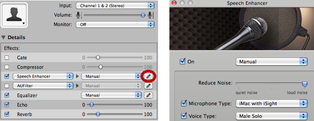
I have some background noise (hiss) when recording my voice audio if both the iMac and the HP laptop are running. I used the GarageBand noise filter to remove some of the background noise:

Noise reduction in GarageBand

Noise reduction in GarageBand

Related blog post: using animated pointers in a narrated PowerPoint presentation.

Posted in learning | Tagged , , , , , | 1 Comment

Prezi presentation software

Is Prezi more useful than PowerPoint?

Testing Prezi on Prezi

Posted in Uncategorized | Leave a comment

Medical Student Bridge Program

I had a chance to observe the University of Arizona Medical Student Bridge Program. We would like to start a similar program at SCNM in order to help “nontraditional” medical students get ready for their first quarter of medical school.

Since I never went to medical school, I am not very good at providing medical students with advice for how to survive in medical school. I’ve long been amazed by the vast amount of information that medical students are able to learn and apply. I benefitted from seeing how the U of A Bridge program advises its new students about how to remember, integrate and use information from their basic science classes.

One of the U of A students that I spoke to said that he was a mathematics major as an undergraduate and that he had never really needed to study previously since mathematics came naturally to him. I had a similar experience in my schooling, being able to select and attend only the classes that I was interested in and not being subjected to a flood of information.

I’ve long had an interest in learning and memory, so I’m familiar with the importance of repetition and the idea that we can all benefit from thinking about information while using multiple sensory modalities. When I was in school I never found it useful to study with someone else. There always seemed to be a temptation to goof off and not study or one person would understand the material and be bored while the other might just be bored with the topic and not understand it.

In the U of A Bridge Program there is an emphasis on the students working together with a partner to either preview material before a lecture or review the information from a lecture that has already been attended. I can see that if medical students follow this approach then they will experience useful repetition while they read, listen to, write and talk about the subject matter. Yet another learning option provided in the U of A medical school is practice exams. Rather than being provided with prectice questions in the form of question sets at the end of textbook chapters or in a board review book, all of the U of A exams and practice questions are online as part of their U of A medical school learning resources.

I’d be interested to hear from SCNM students who can answer this question: What do you now know about how to survive in medical school that you wish you had been told before day 1 of the first quarter?


Related reading

Posted in learning | Tagged , | Leave a comment

Functions of dopamine in the basal ganglia

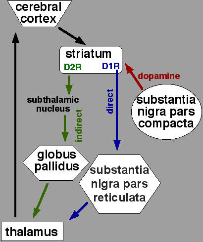
Direct and indirect motor pathways

Direct and indirect motor pathways in the basal ganglia

Parkinson disease can be caused by loss of dapaminergic neurons in the substantia nigra. There are two major populations of neurons in the striatum that respond to dopamine. Their roles in movement disorders are the subjuct of ongoing research.

I previously mentioned the article “Distinct subclasses of medium spiny neurons differentially regulate striatal motor behaviors” which describes experiments in mice that test the current model of how the basal ganglia control movement.

In current models of motor control by the basal ganglia, a distinction is made between a population of type 2 dopamine receptor (D2R) expressing neurons and a population of D1R expressing neurons (see the figure, above right).

By using the gene expression control regions of the D2 and D1 receptors, Bateup et al. were able to selectively disrupt expression of DARPP-32 in either the D2R- or D1R-expressing striatal neurons of mice. DARPP-32 is an important protein for many of the normal actions of dopamine in the brain, including those in the striatum. When DARPP-32 levels were reduced selectively in the D2R-expressing striatal neurons, the mice showed increased locomotor activity. Reduced DARPP-32 in D1R-expressing striatal neurons resulted in reduced basal locomotor activity, consistent with the standard model in which  the direct pathway normally exerts a stimulatory effect on locomotion.

Such “proof of concept” experiments in mice raise hope for future interventions that might allow selective modulation of the direct and indirect pathways in humans. This might help solve problems such as development of dyskinesias following long-term use of L-DOPA or cell transplant therapy in Parkinson disease patients.

Posted in dyskinesia, Huntington disease, L-DOPA, Parkinson disease | Tagged , , , | 1 Comment

Journal Club

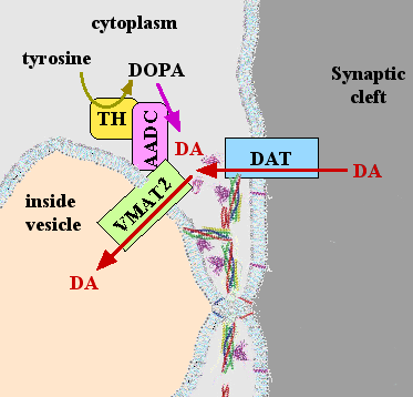
Dopamine transport

Dopamine transport proteins VMAT2 and DAT. (public domain illustration by John Schmidt)

I’m trying to establish a more formal system for a “journal club”, but for now we can use this blog. [Update, Spring 2012, see this journal club page.]

This blog post starts the “journal club”. “Journal club” means that participants will each pick a topic and find a related journal article.

Here I introduce the article called, “Physical and Functional Interaction between the Dopamine Transporter and the Synaptic Vesicle protein Synaptogyrin-3“.

This journal article is medically important because many pharmacological agents alter dopamine (DA) transport and brain function. Methamphetamine is a major drug of abuse and it disrupts normal dopamine transport, but the exact mechanism is still mysterious. As shown in the diagram, two major dopamine transport proteins are DAT and VMAT2. DAT normally allows for re-uptake of dopamine from the synaptic cleft. VMAT2 is important for transport of dopamine from the cytoplasm into synaptic vesicles.

Egaña et al. report some experiments suggesting that there might be a complex of physically interacting proteins including DAT, synaptogyrin-3, and VMAT2. Synaptogyrin-3 is a synaptic vesicle protein. It was also recently suggested that VMAT2 forms a protein complex with tyrosine hydroxylase (TH) and aromatic amino acid decarboxylase (AADC, also called DOPA-decarboxylase).

Why is this medically important? Amphetamines cause depletion of dopamine from synaptic vesicles. If DAT and VMAT2 are physically linked then it might be that amphetamine binding to cell surface transporters like DAT can alter the function of VMAT2. VMAT2 seems to be the binding site for chemicals such as lobelane that can inhibit methamphetamine-evoked DA release from neurons. There might be a previously unrecognized macromolecular complex of proteins linking cell surface transport proteins such as DAT to VMAT2. If such a complex of linked dopamine transport proteins does exist, study of how DAT and VMAT2 interact might help us better understand medical conditions such as Parkinson disease, dystonia, depression, schizophrenia, attention-deficit hyperactivity disorder (ADHD), Tourette syndrome, and drug addiction.

Note: The current conventional thinking about how methamphetamine alters dopamine storage and transport does not involve any physical link between the cell surface DAT and synaptic vesicles.

Related reading.  A Receptor Mechanism for Methamphetamine Action in Dopamine Transporter Regulation in Brain

Is Parkinson’s disease a vesicular dopamine storage disorder? J. Neurosci. 2014 June

Comments are welcome here about the article “Physical and Functional Interaction between the Dopamine Transporter and the Synaptic Vesicle protein Synaptogyrin-3” and dopamine transport in the brain.

If you want to participate in the “journal club”,  find a recently published journal article and prepare a brief written account of why the article is of interest to you. Contact me and I can make it possible for you to post and share your contribution to the “journal club” here in blog format.

Posted in L-DOPA, membrane transport | Tagged , , | 5 Comments

Team-Based Learning

Planet Hollywood

Team-Based Learning Collaborative meeting March 2-4 2011

The Conference on Team-Based Learning in Higher Education was held March 2-4, 2011.

On Tuesday there was an introductory session about the fundamentals of what constitutes a Team-Based Learning (TBL) session. I learned that it is a normal part of many TBL sessions to take some time after the Readiness Assessment Testing (RAT) for a “mini lecture” during which topics that were not well understood  by the students are reviewed. Alternatively, some instructors allow students to write questions about the Readiness Assessment on a whiteboard. Other students in the class can then provide answers to those questions. The instructor can be a facilitator in helping the students work together to learn the material without giving a lecture during the TBL session.

Many institutions are using TBL with a great emphasis on a need for students to read before the TBL session. In many cases, there is NO lecture before a TBL session, only assigned reading. This often makes it more important to have a “mini lecture” after the Readiness Assessment…such “mini lectures” might be the only lectures in the course!

Many medical schools are using TBL for advanced medical students and there is an attempt to design questions that do not have a clear best answer. The objective is to model real-world situations where a physician tries to help a patient under conditions where there is incomplete or misleading diagnostic information. In many cases, the goal of the TBL session is as much to stimulate discussion and the development of collaborative problem solving skills as it is to identify the correct answer. In many situations, the application exercises are not even graded and the only goal is to stimulate the development of problem solving skills in a team.

Objectives for TBL sessions are not simply topics that are included in the session. In a medical school setting, TBL objectives are often specific types of problem solving skills that a physician needs to have. For Human Biology, the real objective of TBL sessions might often be something like, “apply basic science knowledge in topic areas A, B and C to understanding of a specific medical condition”. Can students apply their knowledge of the human body to medical problems? Another type of TBL objective might be, “Students read and understand a published journal article and are able to use the research findings reported in that article to guide patient treatment.”

Before attending the Conference on Team-Based Learning, I thought that the main goal of a TBL session was to have learning take place during discussions of application questions by team members. However, many instructors try to create ambiguous application questions that will cause several different teams to answer one question in different ways. Much of the learning then takes place when teams of students argue about their reasons for selecting different answers to the same question.

One of my major concerns about TBL is that even when a team selects the correct answer to an application question, some members of the team might not really understand why it is the correct answer. One way to assess this possibility is to identify students who missed a particular Readiness Assessment Test question and who were on a team that correctly answered a related application question. At the end of the TBL session, can even those students who missed the Readiness Assessment Test question provide an oral justification for the correct answer on the  application question? The class can be warned that students who do poorly on the Readiness Assessment Test will be called on to justify their team’s answers to the application questions. This would provide strong motivation for teams to make sure that all team members understand the answers to the application questions by the end of the class.

One of the interesting research findings discussed at the meeting is the idea that student retention of information might be better when topics are learned during group discussions and team-based problem solving sessions. TBL is relatively new and not much research has been done exploring the benefits of TBL over other strategies for learning. The results of one small study presented at the meeting suggested that lower quartile students might benefit the most from TBL. Each institution probably needs to have its own on-going research to objectively determine that TBL actually has benefits for its students.

An interesting part of the Conference was that it provided me with my first opportunity to take part in TBL sessions as a “student”. The Conference workshops were formatted so as to include workshop participants in TBL sessions. While watching teams of medical students during the past two quarters I have had doubts about the optimal size of teams. My personal experience at the Conference enforced the feeling that I have had from watching teams try to have discussions. I doubt if 7 students if optimal for allowing team discussions. I spoke to many instructors who use smaller teams (4-5). I’d like to hear from students about this issue (team size) and any other ideas for how to improve TBL sessions in Human Biology.

Image credits. Planet Hollywood Las Vegas by Juan David Ruiz (Creative Commons Attribution 2.0 Generic).

Posted in learning, Team-Based Learning | Tagged , , | Leave a comment

Confidence intervals

confidence intervals

Reporting confidence intervals.

Confidence intervals are often used in biomedical research as a tool for describing the statistical significance of findings in a research study. An example of  such a study is “Examining the association between childhood asthma and parent and grandparent asthma status: Implications for Practice“. This study investigated the association between a family history of asthma and the incidence of asthma in a population of children. Specifically, the study tested for a statistically significant association between childhood asthma incidence and having a parent or grandparent who had asthma.

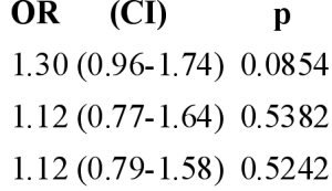
As shown in the figure (above, right), many research reports describe their findings by making use of odds ratios (OR), confidence intervals (CI) and p-values. In this study, children who had a parent with asthma were almost twice as likely (OR = 1.96) to have asthma compared to those children who did not have a parent with asthma. Children who had both a parent and a grandparent with asthma were over four times more likely to have asthma compared to those without a parent or grandparent with asthma (OR = 4.27).

These odds ratios were calculated by first measuring the incidence of asthma in several subgroups of the study population. To calculate an odds ratio, a reference group was defined such as children who did not have either a parent or a grand parent with asthma. The values for OR are calculated as the probability of childhood asthma in a specified subgroup divided by the probability of childhood asthma in a reference group.

How can we know if the odds ratios reported in this study (such as 1.96) are statistically significant? The authors of the study provided both calculated p-values and 95 % confidence intervals for each odds ratio. The greater observed incidence of asthma in children who had a parent with asthma was reported as an odds ratio of 1.96 and a 95 % confidence interval of 1.26-3.06 and a p-value of 0.0027. The 95 % confidence interval indicates a range of odds ratio values that might occur by chance. If the 95 % confidence interval includes 1.0 then there is a greater than 1 in 20 chance that random variation in outcome incidence among the two compared study subgroups can account for the observed difference in incidence between the test group and the reference group.

For p-values, a value less than 0.05 indicates that there is less than an estimated 1 in 20 chance that random variation in outcome incidence among the study subgroups can account for the observed difference in incidence between a test group and the reference group. For the “had a parent with asthma” test group, a p-value of 0.0027 was reported, so there was a greater than 95 % calculated probability that the 1.96 odds ratio indicates a significant difference in asthma incidence between the “had a parent with asthma” test group and the reference group of children who did not have a parent with asthma.

In this study, an example of an odds ratio that was not found to be statistically significant was 1.12 for children who had a parent (mother or father) who smoked (see the  table, above). The reported p-values were greater than 0.05 and the 95 % confidence intervals included 1.0.

The larger an odds ratio and the further the bottom of the 95 % confidence interval is from 1.0 the more likely that an observer odds ratio indicates a statistically significant difference in asthma incidence in the test group. For the test group of children with both a parent and a grandparent with asthma the odds ratio was 4.27, the 95 % confidence interval was 2.39-7.65. The calculated p-value was less than 0.0001. The smaller the p-value the more likely that a study finding is not due to random variations and actually indicated a real difference between two subgroups in the study.

Posted in statistics | Tagged , , | Leave a comment

Platelet activation

adhesion of platelets to collagen-coated surfaces

platelet adhesion

A previous blog post concerned the protein tissue factor and how rupture of blood vessel walls initiates fibrin clot formation. Platelets are cell fragments in the blood that normally lead a solitary existence, but they can quickly attach to damaged blood vessels and start participating in blood clot formation. Adherence of platelets at sites of blood vessel rupture usually initiates changes in platelet behavior that allow platelets to fully participate in hemostasis. In particular, activated platelets help support fibrin clot formation and they release chemical signals that stimulate vascular smooth muscle contraction. These two responses to blood vessel wall damage limit blood loss.

Normally, platelets exist in a special environment within the blood. However, extracellular matrix proteins such as collagen are always nearby, just on the other side of the endothelial lining that normally forms the inner surface of blood vessels. Platelets have receptors for extracellular matrix components such as collagen (1). The images (to the right) show platelets that have attached to a solid substrate that had been coated with collagen.

Along with collagen, von Willebrand factor plays an important role in the adhesion of platelets to ruptured blood vessels (2). The binding of platelet receptor proteins to collagen and von Willebrand factor activates signal transduction pathways that change the behavior of platelets, initiating the process of “platelet activation”. Thrombin production is stimulated by blood vessel wall damage (see this earlier discussion), and thrombin also acts on platelets to promote their activation.

Platelet activation involves several important changes in platelet function that contribute to hemostasis:

1. Activated platelets have increased activity of the enzyme cyclooxygenase and produce the signaling molecule thromboxane A2 (3). Thromboxane A2 has two major effects on hemostasis. It both helps to further activate platelets (4) and it acts on vascular smooth muscle to promote vasoconstriction (5), which limits blood flow through damaged blood vessels .

2. Activated platelets serve as an assembly site for proteins that promote the production of thrombin and fibrin clot formation (recent concise description).

3. Activated platelets release the contents of their secretory vesicles (6) including ADP and von Willebrand factor which promote further platelet activation and aggregation.

4. Activated platelets aggregate (7, 8) by means of their receptors for von Willebrand factor and fibrinogen. In the context of hemostasis, platelet aggregation occurs in parallel with fibrin clot formation.

The parallel production of thrombin and activated platelets involves several positive feedback processes that result in rapid production of a platelet-rich fibrin clot at locations of blood vessel rupture.

Image credits. These images of platelets adhering to a collagen-coated surface are from Figure 3 in Efficient Inhibition of Collagen-Induced Platelet Activation and Adhesion by LAIR-2, a Soluble Ig-Like Receptor Family Member, © copyright 2010 Lenting et al., an open-access article distributed under the terms of the Creative Commons Attribution License.

Posted in hemostasis | Tagged , | Leave a comment